Breast cancer screening saves lives.
But choosing the right test often causes confusion.
Two common options: mammography and ultrasound.
A newer option: 3D mammography.
Each serves a different purpose.
Understanding the differences helps you make better decisions.
What Is Mammography and How It Works
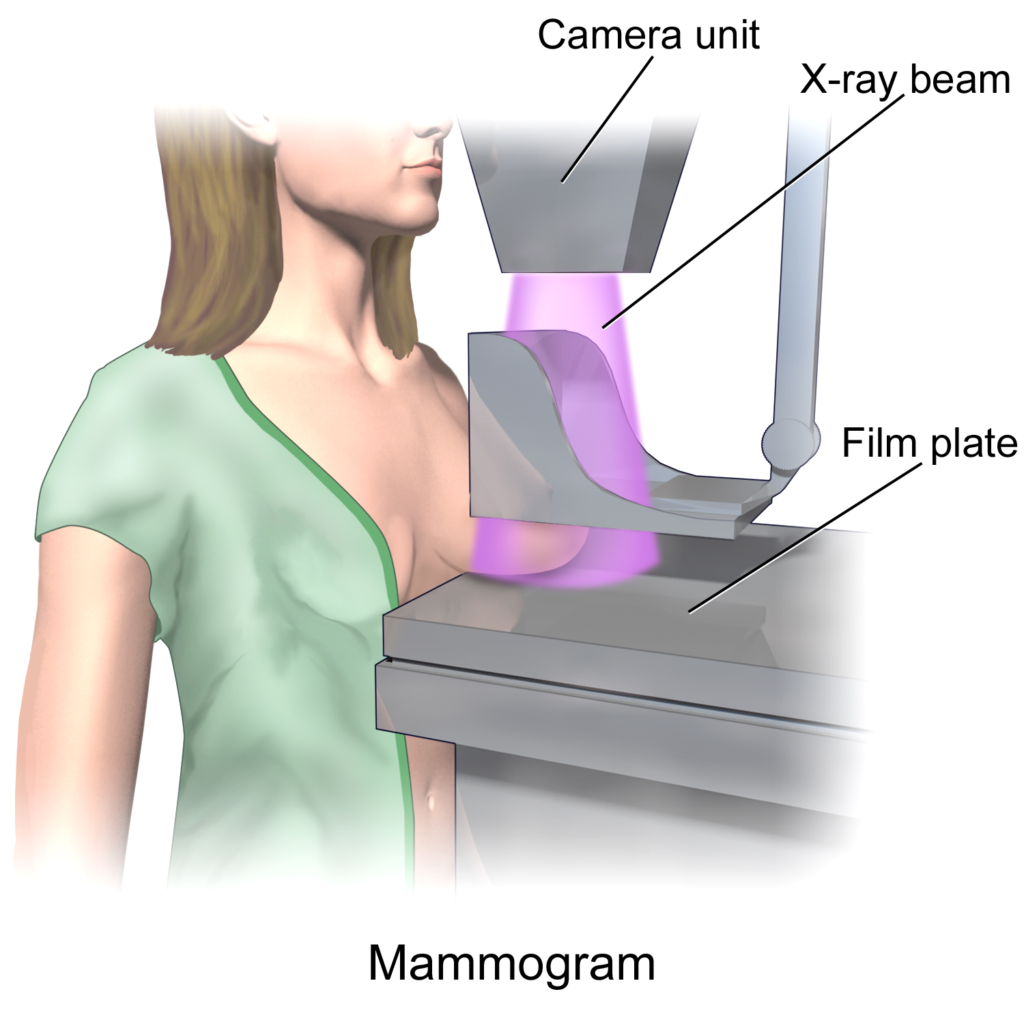
Mammography is an X-ray of breast tissue.
It detects abnormal growths before symptoms appear.
The process involves compressing the breast between two plates.
This improves image clarity and reduces radiation dose.
Mammograms are widely used for routine screening.
They can detect tiny calcium deposits, often an early cancer sign.
Doctors recommend mammography as the first-line screening tool.
Especially for women above 40.
It is fast.
It is standardized.
It has proven survival benefits.
However, its accuracy reduces in dense breast tissue.
That’s where additional imaging may be needed.
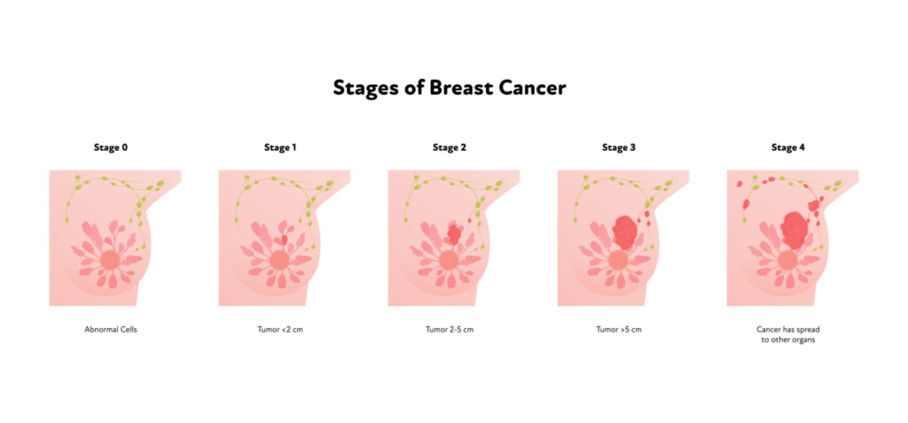
Types of Mammography (2D vs 3D)
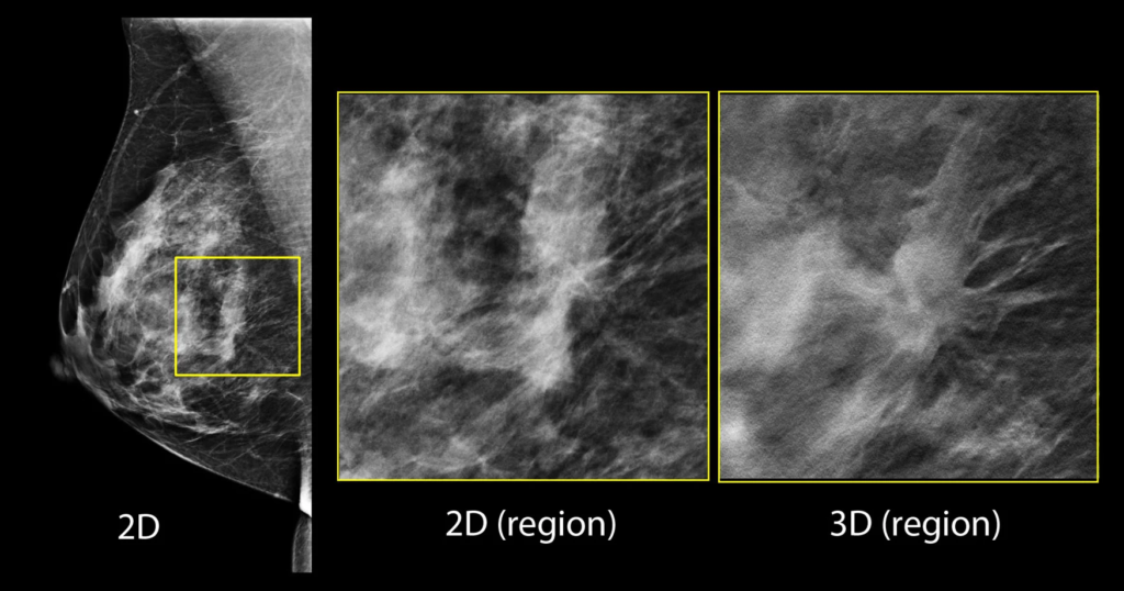
2D mammography captures flat images.
It can miss small tumors hidden in dense tissue.
3D mammography (tomosynthesis) creates layered images.
It allows doctors to examine the breast slice by slice.
This reduces overlapping tissue issues.
Detection improves, especially for small cancers.
False positives also decrease.
Meaning fewer unnecessary biopsies.
When Doctors Recommend Mammography
- Routine screening after age 40
- Family history of breast cancer
- Detecting early-stage tumors
- Follow-up after abnormal findings
It is often the first test, not the only one.
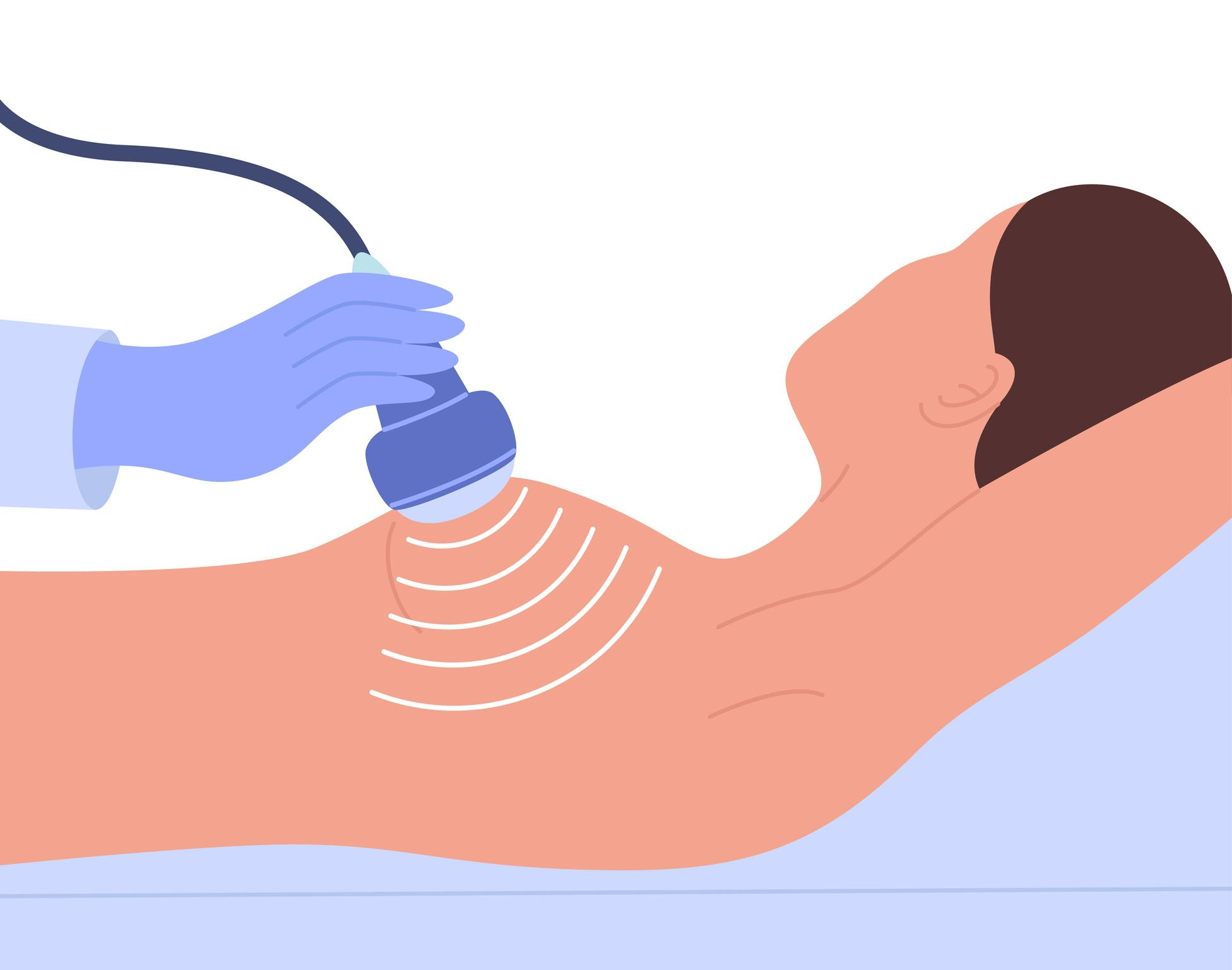
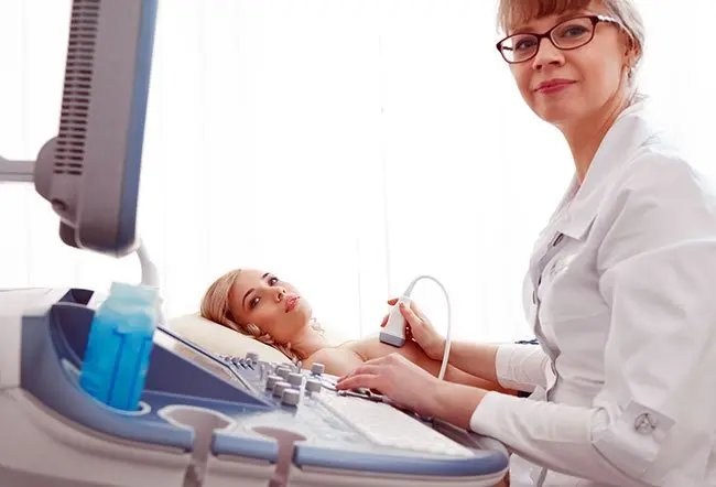
What Is Breast Ultrasound and How It Works
Ultrasound uses sound waves, not radiation.
It creates real-time images of breast tissue.
A gel is applied.
A handheld probe scans the breast.
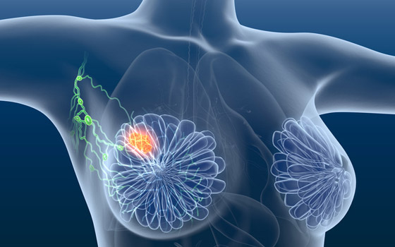
It helps differentiate between solid masses and fluid-filled cysts.
This makes it useful for further evaluation.
Ultrasound is often used after a mammogram.
Not usually as a primary screening tool.
It is painless.
It is safe during pregnancy.
When Ultrasound Is Preferred
- Dense breast tissue
- Younger women under 40
- Pregnant patients
- Evaluating lumps found in physical exams
It complements mammography, not replaces it.
Limitations of Ultrasound
Ultrasound cannot detect microcalcifications well.
These are early signs of cancer.
It also depends heavily on operator skill.
Results may vary.
It may miss very small tumors.
Hence, not ideal for screening alone.
Mammography vs Ultrasound: Key Differences
Both tests serve different roles.
Mammography is for screening.
Ultrasound is for evaluation.
Mammography detects early cancer signs.
Ultrasound provides additional clarity.
Using both together improves diagnosis accuracy.

Accuracy Comparison
Mammography detects early-stage cancer better.
Especially microcalcifications.
Ultrasound is better for characterizing lumps.
It identifies cyst vs solid mass.
For dense breasts, combined imaging works best.
No single test is perfect.
Safety and Radiation
Mammography uses low-dose radiation.
The risk is minimal.
Ultrasound uses no radiation.
It is completely safe.
For frequent screening, benefits outweigh risks.

3D Mammography: Is It More Accurate?
3D mammography improves detection rates.
Especially in dense breast tissue.
It captures multiple images from different angles.
These are reconstructed into thin slices.
Doctors can examine each layer clearly.
This reduces missed cancers.
Studies show higher detection rates.
Also fewer false alarms.
It is becoming the new standard in many hospitals.
Benefits of 3D Imaging
- Better detection in dense breasts
- Reduced false positives
- Clearer tumor visualization
- Fewer repeat tests
It improves confidence in diagnosis.
Who Should Consider 3D Mammography
- Women with dense breast tissue
- Family history of breast cancer
- Previous abnormal mammograms
- High-risk individuals
It offers better screening precision.
Which Test Should You Choose?
The choice depends on multiple factors.
Age.
Risk level.
Breast density.
Doctors often combine methods for accuracy.
Based on Age and Risk
- Under 40: Ultrasound preferred
- 40+: Mammography recommended
- High risk: Combination approach
Screening frequency also varies.
Based on Breast Density
Dense breasts reduce mammogram accuracy.
In such cases:
- Add ultrasound
- Consider 3D mammography
Personalized screening works best.
Final Thoughts
There is no single “better” test.
Mammography is essential for screening.
Ultrasound supports diagnosis.
3D mammography improves accuracy.
The best approach is combined and personalized.
Early detection remains the priority.
Consult a qualified specialist.
Choose screening based on your risk profile.
Suggested Internal Links
- Early signs of breast cancer
- Breast biopsy explained
- Lifestyle changes to reduce cancer risk
Suggested External Links
- WHO breast cancer guidelines
- Indian Cancer Society
- CDC breast screening recommendations
Consult Dr. Pollob Saha – Expert Laparoscopic Surgeon in Kolkata
Dr. Pollob Saha is a highly trusted general and laparoscopic surgeon in Kolkata, known for his skilled hands and patient-first approach. From initial consultation to full recovery, he ensures comprehensive care and support for every patient.
📍 Website: https://drpallabsahasurgeonkolkata.in/
📧 Email: drpallabsaha20@gmail.com
📞 Phone: (+91) 9830088321
Book your consultation today and prepare for your laparoscopic surgery with complete confidence!