With my 18 years of experience as a doctor, I remember when planning a breast cancer operation meant sitting with a set of flat images on a lightbox and making your best judgment.
You’d look at the mammogram. You’d study the ultrasound. You’d read the MRI report. Then, using your experience and clinical knowledge, you would try to visualize what the breast looked like on the inside-where the tumour was located, how deep it extended and what surrounding structures might be involved. It was like building a mental picture from two-dimensional clues and hoping that picture matched reality. Sometimes it did. Sometimes it didn’t and you only discovered the difference once you were already in the operating theatre.
Today, the way we approach 3D imaging breast cancer surgery has transformed that process. Surgeons can now view detailed three-dimensional representations of the breast and tumour before making a single incision, allowing for more accurate planning and better-informed surgical decisions.
That’s changed now. Not completely, but definitely in a meaningful way. And 3D imaging is one of the biggest reasons why.
The Problem with Flat Images
Here’s something that took me a while to articulate clearly to patients: breast tissue is not flat. Neither is a tumour.
When we look at a conventional mammogram or a standard MRI slice, we’re seeing a cross-section-a single plane through a three-dimensional structure. It’s a bit like trying to understand a building by looking at one floor plan. You get information, but you’re missing the whole picture.
Tumours sit at varying depths. They extend in different directions. They have relationships with nearby structures like the nipple, the underlying muscle and the lymph nodes-relationships that matter enormously when you’re planning how to operate. A flat image can suggest those relationships, but it can’t show them to you the way a 3D model can.
This isn’t a criticism of the radiologists or the technology that came before. Those tools saved lives and they still do. But when I’m planning an operation, I want the most complete spatial picture possible. Effective breast cancer surgical planning depends on understanding exactly where the tumour sits and how far it extends within the breast. Two-dimensional imaging, used alone, leaves gaps that 3D imaging helps close.
So What Does 3D Imaging Actually Mean?
For most people when they hear the term 3D imaging , they imagine some complex futuristic process like the one we see on sci-fi movies and series but the reality is less dramatic but more useful.
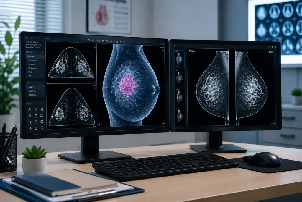
What these technologies share is a simple principle: they take raw imaging data and reconstruct it into something you can navigate in three dimensions. Instead of reading a shadow on film, you’re working with a model you can rotate, slice and measure from any angle you need. Before a single incision is made.
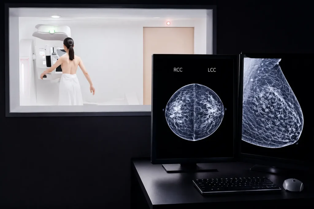
Digital breast tomosynthesis is probably the most familiar example , it’s what clinics mean when they advertise 3D mammography. Regular mammography takes one compressed image. Tomosynthesis takes many images from slightly different angles and assembles them into thin cross-sectional slices, like the pages of a book. You work through them one by one.
The practical benefit is significant.
Dense breast tissue which is genuinely common and genuinely problematic for standard imaging – becomes far more readable. Small cancers that were hiding behind overlapping tissue become visible. Fewer false positives means fewer patients going through unnecessary biopsies, which is not a small thing.
The Margin Problem and Why It Matters So Much
If I had to name one thing that keeps breast surgeons up at night, it’s margins.
A clear margin – a negative margin means the tumour has been removed with a rim of healthy tissue around it. So no cancer cells are present at the edge of the specimen. When that’s what the pathology shows, the patient and the doctor can both breathe a little easier.
When the margin comes back positive, it means cancer cells were found at the very edge of the tissue that was removed. Which means there’s a real chance some were left behind. And that almost always means going back to the theatre.
Another operation. Another anaesthetic. More recovery time. And the psychological experience of being told that the surgery didn’t quite get everything, that’s genuinely hard for patients to hear.
Positive margins aren’t always about surgical technique. More often, they come down to preoperative information. The surgeon didn’t fully know the extent of the tumour before starting. The map was incomplete.
And exactly here 3D imaging changes that. Studies consistently show higher rates of clear margins when surgeons use volumetric preoperative imaging. When you’ve essentially rehearsed the resection in a digital environment before the operation when you understand where the tumour ends in all three dimensions, you approach the case with a level of precision that’s genuinely hard to achieve any other way.
It’s like this: the better your map, the fewer wrong turns.
Oncoplastic Surgery: Thinking About What Comes After
Removing cancer is one goal. What the breast looks like and feels like afterwards is another and one that patients care about enormously, even if they sometimes feel they shouldn’t say so.
Oncoplastic breast surgery tries to address both goals at once. We’re not just removing the tumour; we’re thinking simultaneously about how to preserve or reconstruct the breast’s shape, symmetry and volume. Done well, it means patients don’t have to choose between adequate cancer treatment and a result they can live with comfortably.
To plan that properly, the doctor need to know precisely how much tissue is coming out and exactly where the defect will be created. 3D surface imaging systems use structured light to create a digital model of the breast’s external contour which allow us to simulate different
During the Operation Itself
I want to be honest about something: even a beautifully detailed preoperative plan meets reality when you start operating the patient in the theatre.
Breast tissue shifts when a patient is repositioned on the table. Something that looked a certain way on MRI can present differently under direct examination. A tumour you were confident about suddenly feels less clear-cut.
Intraoperative ultrasound with real-time 3D reconstruction addresses this. During the operation, the imaging continues. You can re-evaluate tumour position, check margin adequacy as you go and adjust based on what you’re actually seeing rather than what you anticipated seeing.
The Conversation Before Surgery
This is something I want to talk about separately, because I think it gets undervalued.
When a patient comes to see me before a breast cancer operation, I have an obligation beyond the technical. I need to make sure she actually understands what we’re planning to do and why. Not as a legal formality but as a basic matter of respect.
The problem is that surgical margins and resection volumes and oncoplastic reconstruction are not layman concepts. Explaining them well, to someone who is frightened and processing a diagnosis, is genuinely difficult.
A 3D model helps. When I can show someone a volumetric representation of their breast – tumour visible, planned excision marked ,the conversation stops being abstract. They can see it. They can point to it and ask questions. They understand, in a tangible way, what is going to happen and what we’re trying to achieve.
Patients who go into surgery with that kind of understanding seem to recover differently. They’re less blindsided by what they experience. They know what to expect, more or less. The informed consent conversation stops being a form to sign and becomes an actual exchange between two people.
That’s what I think good surgical care should look like.
Is This Available in Kolkata?
Patients ask me this regularly and I understand why. There’s sometimes an assumption that the more sophisticated the technology, the less accessible it is.
The honest answer is: more than you might think.
Digital breast tomosynthesis is available at several major diagnostic centres in Kolkata. MRI-based 3D reconstruction is offered at tertiary oncology facilities. The gap between what’s accessible here and what’s considered standard at leading centres internationally has narrowed substantially and it continues to narrow.
If you’re planning breast cancer surgery in Kolkata, ask your surgical team directly about imaging. What has been done? What did it show about the extent of the tumour? How is that information shaping the operative plan? These are completely reasonable questions. You’re not being difficult by asking them. You’re being a good advocate for yourself.
Where Things Are Heading
AI tools are being integrated with volumetric imaging system in ways that are beginning to identify radiological features associated with tumour behaviour, predict margin adequacy before the operation and highlight cases that warrant more thorough workup. These tools are still maturing.
Robotic-assisted breast surgery, still in early adoption, is being designed to interface with preoperative 3D data directly. The digital plan becomes the parameters the robotic system works within. The distance between planning and execution shrinks further.
Whether all of this delivers on its promise remains to be seen. But the direction is clear.
A Final Word
3D imaging isn’t magic. It doesn’t replace surgical judgment and it doesn’t eliminate uncertainty. But it gives that judgment something better to work with.
It improves the odds of getting clear margins. It supports more thoughtful oncoplastic planning. It makes the patient conversation richer and more real. And it keeps improving.
If surgery is being planned for you, or someone you care about then ask about imaging. Ask how the tumour has been mapped. Ask what the plan is for margins. These questions matter and the answers will tell you a lot about the care you’re receiving.
Early diagnosis and expert, thoughtful surgical care are still the most powerful tools we have. That hasn’t changed. But the tools that support them keep getting better.
Frequently Asked Questions
1. What is 3D imaging in breast cancer surgery?
3D imaging uses advanced technologies such as digital breast tomosynthesis, MRI-based reconstruction and 3D ultrasound to create a detailed three-dimensional view of the breast and tumour. This helps surgeons plan procedures more accurately before surgery.
2. How does 3D imaging improve breast cancer surgery outcomes?
By providing a clearer understanding of the tumour’s size, location and relationship to surrounding tissues, 3D imaging helps surgeons achieve more precise tumour removal, improve margin clearance and reduce the likelihood of additional surgeries.
3. Does 3D imaging reduce the need for repeat breast cancer surgery?
In many cases, yes. Better preoperative planning can help surgeons remove the entire tumour with clear margins during the first operation, lowering the chances of needing a second procedure.
4. Is 3D mammography better than a standard mammogram?
3D mammography, also known as digital breast tomosynthesis, can detect certain breast cancers more effectively, especially in women with dense breast tissue. It may also reduce false-positive findings and unnecessary follow-up tests.
5. Can 3D imaging help preserve the appearance of the breast after surgery?
Yes. 3D imaging supports oncoplastic breast surgery by helping surgeons estimate how much tissue needs to be removed and plan reconstruction techniques that maintain breast shape and symmetry whenever possible.
📞 Phone: (+91) 9830088321
🌐 Website: https://drpallabsahasurgeonkolkata.in/
📧 Email: drpallabsaha20@gmail.com